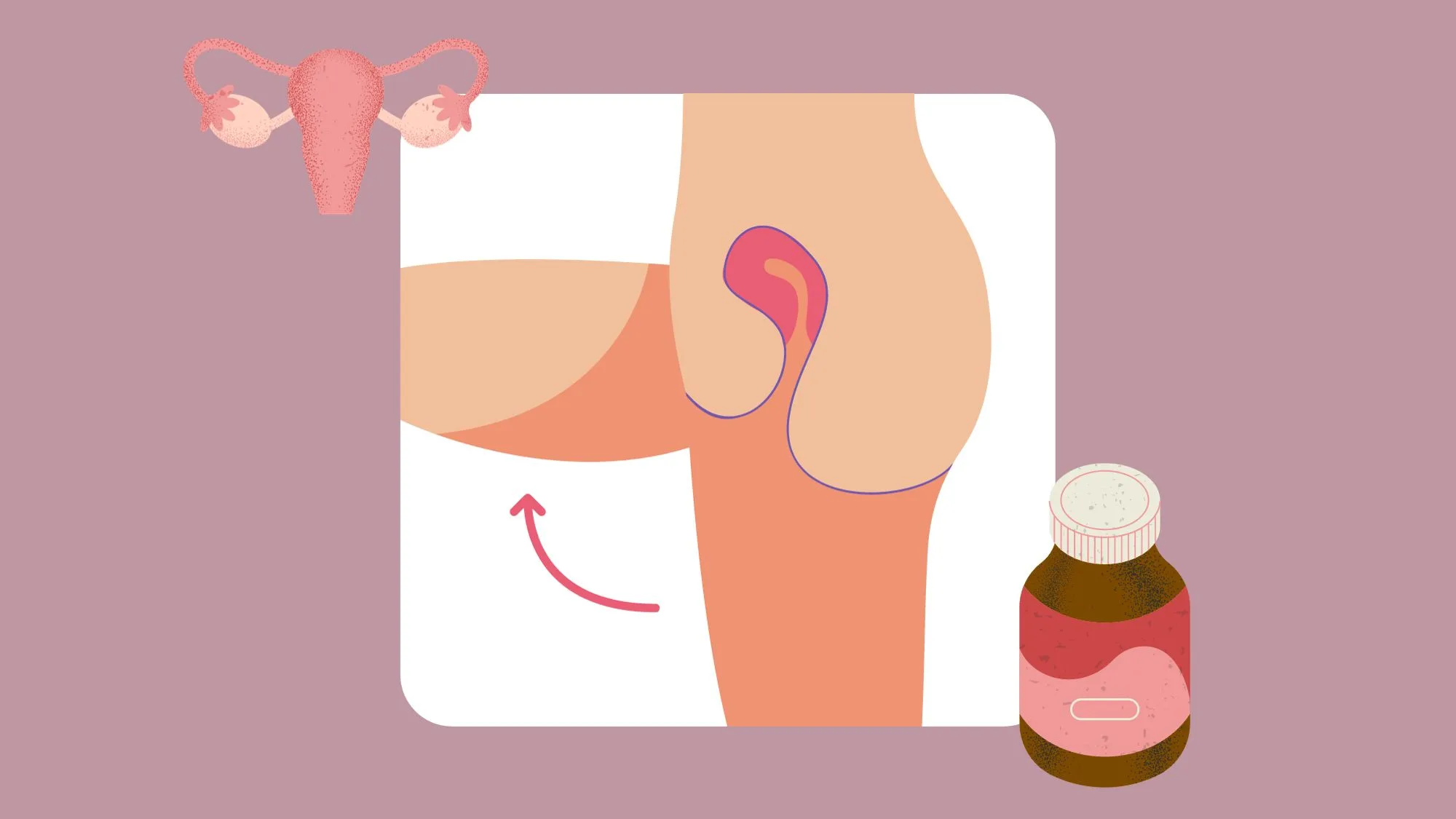
Uterine inversion is a rare but serious complication. It occurs when the uterus turns inside out after childbirth or in non-pregnant individuals. This article aims to provide a comprehensive understanding of uterine inversion. Including its causes, symptoms, diagnostic procedures, treatment options, prevention strategies, and the emotional and psychological aspects of living with this condition.[1][2][3][4][5]
What is Uterine Inversion?
Uterine inversion is a condition in which the uterus, or womb, collapses and turns inside out. This happens when the top of the uterus descends into the uterine cavity or even protrudes outside the body. The inversion can be partial. Where only a portion of the uterine wall turns inward, or complete. Where the entire uterus is inverted. Regardless of the type, uterine inversion requires immediate medical attention.
Definition and Overview of Uterine Inversion
Uterine inversion refers to the folding or inversion of the uterus. Resulting in a protrusion or complete turning inside out of the organ. This condition is considered a medical emergency due to the potential risk of severe hemorrhage and infection. Uterine inversion can occur either during childbirth or in non-pregnant individuals.
The Anatomy of a Normal Uterus vs an Inverted Uterus
A normal uterus is a pear-shaped organ located in the pelvis. Which is held in place by ligaments and muscles. In an inverted uterus, the top part of the uterus collapses into the cavity or even extends outside the body. This reversal of the uterine wall can disrupt blood flow. Leading to complications such as excessive bleeding and shock.
When examining the anatomy of a normal uterus, it is important to understand its role in reproduction. The uterus is a vital organ in the female reproductive system. Responsible for nurturing and supporting a developing fetus during pregnancy. It consists of three layers: the innermost layer called the endometrium, the middle layer known as the myometrium, and the outer layer called the perimetrium. These layers work together to create a suitable environment for implantation and fetal growth.
Inverted Uterus Overview
An inverted uterus presents a significant deviation from the normal anatomy. The collapse and turning inside and out of the uterus can occur spontaneously or as a result of various factors. Such as excessive traction during childbirth, uterine tumors, or uterine atony. The severity of the inversion can vary. Ranging from a partial inversion where only a portion of the uterine wall is affected. To a complete inversion where the entire uterus is inverted.
When an inversion occurs, the blood vessels that supply the uterus can become compressed or stretched. Leading to compromised blood flow. This compromised blood flow can result in ischemia. Which is the inadequate supply of oxygen and nutrients to the affected tissues. As a consequence, the woman may experience severe pain, excessive bleeding, and even shock.
It is worth noting that uterine inversion is a rare condition, occurring in approximately 1 in 2,000 to 1 in 10,000 deliveries. However, when it does occur, it is crucial to recognize the signs and symptoms promptly and seek immediate medical attention. Early diagnosis and intervention can significantly improve the chances of a successful outcome and minimize the risk of complications.
This is a serious medical condition characterized by the collapse and turning inside out of the uterus. It can occur during childbirth or in non-pregnant individuals, and it requires immediate medical attention due to the potential risks of severe hemorrhage and infection. Understanding the anatomy of a normal uterus and the deviations seen in an inverted uterus can help healthcare professionals recognize and manage this condition effectively.[6]
Causes of Uterine Inversion
Uterine inversion, a rare but serious condition, can be triggered by a variety of factors, including postpartum complications and non-puerperal causes. Understanding these causes is crucial for early detection and appropriate management.
Postpartum Uterine Inversion
Postpartum uterine inversion occurs after childbirth and is most commonly associated with excessive traction on the umbilical cord, manual removal of the placenta, or fundal pressure during delivery. These actions can cause the uterus to invert and collapse, leading to a potentially life-threatening situation for the mother.
Excessive traction on the umbilical cord, often done in an attempt to speed up the delivery of the placenta, can exert a strong force on the uterus. This force can cause the weakened uterine walls to collapse inward, resulting in uterine inversion. Similarly, manual removal of the placenta, especially when done forcefully or without proper technique, can disrupt the delicate balance within the uterus and lead to inversion.
Fundal pressure, applied by healthcare providers during delivery to facilitate the expulsion of the baby, can also contribute to uterine inversion. In some cases, excessive or prolonged pressure on the fundus can cause the uterus to turn inside out, posing a serious risk to the mother’s health.
Non-puerperal Uterine Inversion
Non-puerperal uterine inversion refers to uterine inversion that occurs in non-pregnant individuals. While rare, this condition can still have significant consequences and requires prompt medical attention.
One of the primary non-puerperal causes of uterine inversion is the presence of uterine fibroids. These benign tumors, which develop within the muscular walls of the uterus, can distort the normal anatomy and increase the risk of inversion. The size, location, and number of fibroids can all influence the likelihood of uterine inversion occurring.
Other abnormal growths in the uterus, such as polyps or adenomyosis, can also contribute to non-puerperal uterine inversion. These growths can disrupt the structural integrity of the uterus, making it more susceptible to inversion. Additionally, uterine prolapse, a condition where the uterus descends into the vaginal canal, can increase the risk of inversion, although it is relatively uncommon.
In some cases, excessive force during a medical procedure or trauma can also lead to uterine inversion. This can occur during procedures such as hysteroscopy, dilation and curettage (D&C), or even due to external trauma to the abdomen. It is important for healthcare providers to exercise caution and use appropriate techniques to minimize the risk of uterine inversion in these situations.
Understanding the causes of uterine inversion is crucial for healthcare providers and individuals alike. By recognizing the risk factors and taking appropriate preventive measures, the incidence of uterine inversion can be minimized, ensuring the well-being of mothers and non-pregnant individuals alike.[7][8]
Identifying the Symptoms of Uterine Inversion
Recognizing the symptoms of uterine inversion is crucial for prompt diagnosis and treatment. Uterine inversion is a rare but serious condition where the uterus turns inside out and protrudes through the cervix. This can occur immediately after childbirth or during the postpartum period.
Early Signs and Symptoms
Early signs of uterine inversion might include severe abdominal pain, heavy vaginal bleeding, a sensation of something coming out of the vagina, and a visible mass or lump at the vaginal opening. These symptoms can be alarming and require immediate medical attention. It is important not to ignore any unusual sensations or changes in your body, especially after childbirth.
In some cases, the uterus may also be felt as a soft, spongy mass on abdominal examination. This palpable mass can be an important clue for healthcare providers in diagnosing uterine inversion. However, it is crucial to consult a healthcare professional for an accurate diagnosis, as other conditions can present with similar symptoms.
Long-term Effects and Complications
The long-term effects and complications of uterine inversion can vary depending on the severity and duration of the inversion. Immediate medical intervention is necessary to prevent further complications and ensure the well-being of the patient.
Potential complications of uterine inversion include infection, hemorrhage, shock, infertility, and psychological distress. Infection can occur due to the exposure of the inverted uterus to the vaginal environment, which is rich in bacteria. Hemorrhage, or excessive bleeding, can result from the disruption of blood vessels during the inversion process. This can lead to severe blood loss and potentially life-threatening situations.
Shock, a condition characterized by inadequate blood flow to the body’s organs, can occur as a result of the rapid blood loss and subsequent drop in blood pressure. Infertility may be a consequence of uterine inversion, especially if the condition is not promptly treated. The inversion can cause damage to the uterus and its supporting structures, affecting the ability to conceive and carry a pregnancy to term.
Psychological distress is another potential complication of uterine inversion. The experience of having the uterus turn inside out can be traumatic and emotionally challenging for the individual. It is important to provide appropriate psychological support and counseling to help the patient cope with the psychological impact of the condition.
Recognizing the symptoms is crucial for early diagnosis and prompt treatment. The long-term effects and complications of uterine inversion can be severe, highlighting the importance of immediate medical attention. If you experience any concerning symptoms after childbirth, it is essential to consult a healthcare professional for proper evaluation and management.[9][10][11][12][13]
Diagnostic Procedures for Uterine Inversion
To diagnose uterine inversion accurately, healthcare professionals employ various diagnostic procedures.
Uterine inversion is a rare but potentially life-threatening condition in which the uterus turns inside out and protrudes through the cervix. Prompt diagnosis is crucial to prevent complications and ensure appropriate management.
Physical Examination
A thorough physical examination is the first step in diagnosing uterine inversion. The healthcare provider will assess the patient’s medical history, perform a pelvic examination, and evaluate the vaginal opening for any visible signs of inversion.
During the pelvic examination, the healthcare provider may palpate the uterus to determine its position and consistency. In cases of uterine inversion, the uterus may feel soft and enlarged, and the cervix may be displaced or not palpable.
Additionally, the healthcare provider will assess the patient’s vital signs, including blood pressure and heart rate, to evaluate for signs of shock or hemorrhage, which can occur as a result of uterine inversion.
Imaging Techniques
In cases where the diagnosis remains uncertain or further assessment is required, imaging techniques such as ultrasound, MRI, or CT scans may be utilized. These imaging modalities provide a detailed view of the uterus and surrounding structures, aiding in accurate diagnosis and planning of appropriate treatment.
Ultrasound is often the initial imaging modality of choice due to its accessibility, cost-effectiveness, and ability to provide real-time images. It can help visualize the inverted uterus, assess the extent of the inversion, and identify any associated complications, such as placental retention or uterine tears.
In more complex cases or when additional information is needed, MRI or CT scans may be recommended. These imaging techniques offer a more comprehensive evaluation of the pelvic anatomy, allowing for a detailed assessment of the uterine inversion and its impact on adjacent structures, such as the bladder or intestines.
Furthermore, imaging can help differentiate uterine inversion from other conditions that may present with similar symptoms, such as prolapsed uterus or cervical polyps.
Overall, the combination of physical examination and imaging techniques plays a crucial role in the accurate diagnosis of uterine inversion. Prompt recognition of this condition enables healthcare providers to initiate appropriate management strategies, including manual reduction or surgical intervention, to restore the uterus to its normal position and prevent potential complications.[14][15]
Treatment Options for Uterine Inversion
Immediate medical attention is crucial in managing uterine inversion. The treatment approach depends on the severity of the inversion and the individual’s overall health.
Uterine inversion is a rare but serious condition that occurs when the uterus turns inside out and protrudes through the cervix. It can happen during childbirth or shortly after delivery. Immediate intervention is necessary to prevent further complications and ensure the well-being of the patient.
Immediate Management of Uterine Inversion
For acute cases of uterine inversion, the priority is to restore the uterus to its normal position quickly. This may involve gentle manual repositioning of the uterus under anesthesia, followed by administration of fluids and medications to stabilize the patient’s condition.
The manual repositioning procedure requires skill and expertise to avoid causing additional damage. The healthcare provider carefully manipulates the uterus back into its correct position, ensuring that the blood supply to the organ is not compromised. Anesthesia is administered to minimize pain and discomfort during the procedure.
Surgical Interventions
In some cases, surgical interventions are necessary to correct the uterine inversion. This typically involves a surgical procedure known as a laparotomy, where the uterus is repositioned and any necessary repairs are made. In severe cases, a hysterectomy may be performed to remove the uterus entirely and prevent further complications.
Laparotomy is a major surgical procedure that involves making an incision in the abdomen to access the uterus. The surgeon carefully manipulates the inverted uterus back into its normal position and repairs any damage to the surrounding tissues. This procedure may also involve removing any blood clots or debris that may have accumulated in the uterus.
In severe cases or when the patient’s health is at risk, a hysterectomy may be performed. This involves the removal of the uterus, eliminating the risk of future inversions and potential complications. However, this option is only considered when all other treatment options have been exhausted.
Post-treatment Care and Recovery
Following treatment, the individual will require close monitoring and ongoing care. This may involve the administration of antibiotics to prevent infection, pain management, and counseling on postpartum or non-puerperal care. Regular follow-up examinations are vital to ensure proper healing and address any potential complications.
Recovery from uterine inversion can vary depending on the severity of the condition and the individual’s overall health. The patient needs to rest and avoid strenuous activities during the recovery period. Pain medication may be prescribed to manage any discomfort, and it is essential to follow the healthcare provider’s instructions regarding wound care and medication administration.
Emotional support is also crucial during the recovery process. The experience of uterine inversion can be traumatic, and individuals may benefit from counseling or support groups to help them process their emotions and adjust to any physical changes resulting from the condition or its treatment.
This is a serious medical condition that requires immediate attention and appropriate treatment. The management of uterine inversion involves a combination of manual repositioning, surgical interventions, and post-treatment care to ensure the well-being and recovery of the patient.[16]
Prevention of Uterine Inversion
Uterine inversion, although not always preventable, can be minimized by implementing certain measures and addressing risk factors.
One of the key factors in preventing this kind of health condition is identifying and addressing any underlying risk factors. For example, uterine fibroids, which are noncancerous growths in the uterus, can increase the risk. By monitoring the growth and development of fibroids during pregnancy, healthcare professionals can take appropriate measures to minimize the risk. This may include considering alternative delivery methods or even removing the fibroids before delivery.
In addition to fibroids, other abnormal growths or complications during pregnancy can also contribute to the risk of uterine inversion. Regular prenatal care is crucial in monitoring the overall health of the mother and the baby. Through regular check-ups, healthcare professionals can detect any potential issues early on and take necessary interventions to prevent this health condition.
Risk Factors and How to Minimize Them
Identifying and addressing risk factors such as fibroids, abnormal growths, or complications during pregnancy can help reduce the likelihood of uterine inversion. Adequate prenatal care, monitoring, and interventions can play a significant role in preventing this condition.
Another important aspect of preventing uterine inversion is ensuring proper management of any existing conditions that may increase the risk. For instance, if a woman has a history of uterine inversion in a previous pregnancy, healthcare professionals can closely monitor her during subsequent pregnancies and take necessary precautions to minimize the risk.
Furthermore, maintaining a healthy lifestyle during pregnancy can also contribute to the prevention. This includes eating a balanced diet, engaging in regular physical activity as advised by healthcare professionals, and avoiding harmful substances such as tobacco and alcohol.
Role of Regular Check-ups in Prevention
Regular check-ups with healthcare professionals are essential for early detection and management of any underlying conditions that may increase the risk of uterine inversion. Timely interventions, such as the removal of abnormal growths or appropriate delivery methods, can help prevent uterine inversion.
During these check-ups, healthcare professionals will monitor the growth and development of the uterus, as well as assess the overall health of the mother and the baby. This allows them to identify any potential risk factors and take necessary actions to minimize the risk of uterine inversion.
In addition to physical examinations, regular check-ups also provide an opportunity for healthcare professionals to educate pregnant women about the signs and symptoms. By being aware of these indicators, women can seek immediate medical attention if they experience any unusual symptoms, thus enabling early intervention and prevention of uterine inversion.
While this cannot always be prevented, taking proactive measures and addressing risk factors can significantly minimize the risk. Regular prenatal care, monitoring, and interventions are crucial in preventing this condition and ensuring the well-being of both the mother and the baby.[17][18]
Living with Uterine Inversion
Living with this condition involves both physical and emotional aspects that require support and understanding.
It is a rare but serious condition where the uterus turns inside out and protrudes through the cervix. This can occur during childbirth, often as a result of excessive traction on the umbilical cord or improper management of the third stage of labor. The physical consequences of uterine inversion can be significant, requiring immediate medical attention and intervention.
However, the impact of this health condition extends beyond the physical realm. A diagnosis can be emotionally challenging for individuals and their families. The sudden and unexpected nature of this condition can lead to feelings of shock, fear, and uncertainty. It is important to provide emotional support, counseling, and resources to help cope with the physical and psychological aspects of living with this condition.
Open communication plays a vital role in the emotional healing process. Creating a safe space for individuals to express their fears, concerns, and emotions can help alleviate the psychological burden associated with this condition. Support groups, both in-person and online, can provide a sense of community and understanding. Sharing experiences, seeking advice, and finding support from others who have gone through similar situations can be immensely comforting.
Support and Resources for Affected Individuals
Various support networks, online communities, and organizations offer valuable resources, information, and assistance for individuals living with uterine inversion. These platforms provide a space for sharing experiences, seeking advice, and finding support from others who have gone through similar situations.
Additionally, healthcare professionals play a crucial role in supporting individuals with uterine inversion. They can provide guidance on managing physical symptoms, offer information on treatment options, and address any concerns or questions that may arise. Collaborating with a multidisciplinary team, including obstetricians, gynecologists, psychologists, and social workers, can ensure comprehensive care and support for affected individuals.
By recognizing the causes, identifying the symptoms, seeking timely diagnosis, and accessing appropriate treatment options, affected individuals can receive the necessary care and support to navigate this complex condition. Through education, prevention, and emotional support, we can work towards a better understanding and management of your health condition.
Navigating Complexities Using The CareClinic App
With features designed to track symptoms, monitor treatment progress, and log medication schedules, CareClinic provides a comprehensive platform to support your recovery and well-being. By consistently using the app to record your physical and emotional health, you can gain insights into the effectiveness of your treatment plan and make informed decisions with your healthcare team. The app’s reminder system ensures you stay on top of medication and follow-up appointments. While the diary feature allows for reflection on your emotional state. For a proactive approach to managing symptoms and enhancing your health outcomes, Install the CareClinic App today.[19][20]
References
- “Uterine Inversion – Gynecology and Obstetrics – MSD Manual Professional Edition”. https://www.msdmanuals.com/professional/gynecology-and-obstetrics/intrapartum-complications/uterine-inversion
- “Uterine Inversion (Inverted Uterus): Causes & Treatment”. https://my.clevelandclinic.org/health/diseases/22326-uterine-inversion
- “Management of Nonpuerperal Uterine Inversion Using a Combined Vaginal and Abdominal Approach – PMC”. https://pmc.ncbi.nlm.nih.gov/articles/PMC7794039/
- “Uterine inversion | Better Health Channel”. https://www.betterhealth.vic.gov.au/health/conditionsandtreatments/uterine-inversion
- “An Inverted Uterus And Psychological Trauma – Vesna's Story | Birth Trauma Australia”. https://birthtrauma.org.au/an-inverted-uterus-and-psychological-trauma-vesnas-story/
- “Risks and consequences of puerperal uterine inversion in the United States, 2004 through 2013 – PubMed”. https://pubmed.ncbi.nlm.nih.gov/28522320/
- “Characteristics and Outcome in Non-Puerperal Uterine Inversion – PMC”. https://pmc.ncbi.nlm.nih.gov/articles/PMC7971731/
- “Non-puerperal Uterine Inversion with endometrial polyps in an 11-year-old girl: A Case Report. – Journal of Pediatric and Adolescent Gynecology”. https://www.jpagonline.org/article/S1083-3188%2821%2900295-3/fulltext
- “Uterine Inversion – StatPearls – NCBI Bookshelf”. https://www.ncbi.nlm.nih.gov/sites/books/NBK525971/
- “Uterine Inversion – Dermatology Advisor”. https://www.dermatologyadvisor.com/home/decision-support-in-medicine/obstetrics-and-gynecology/uterine-inversion/
- “Uterine Inversion and Uterine Rupture | Anesthesia Key”. https://test.aneskey.com/uterine-inversion-and-uterine-rupture/
- “Uterine Inversion: Diagnosis, Treatment, and its Effects on Future Pregnancies – Mama Net”. https://mamacliqs.com/uterine-inversion-diagnosis-treatment-and-its-effects-on-future-pregnancies/
- “What Is Uterine Inversion? – Klarity Health Library”. https://my.klarity.health/what-is-uterine-inversion/
- “Uterine Inversion and Uterine Rupture | Obgyn Key”. https://obgynkey.com/uterine-inversion-and-uterine-rupture/
- “Uterine Inversion | RadioGraphics”. https://pubs.rsna.org/radiographics/doi/full/10.1148/rg.230004
- “Uterine Inversion | Treatment & Management | Point of Care”. https://www.statpearls.com/point-of-care/30895
- “Contemporary Management of Fibroids in Pregnancy – PMC”. https://pmc.ncbi.nlm.nih.gov/articles/PMC2876319/
- “Uterine Inversion: A Review of a Life-Threatening Obstetrical Emergency – PubMed”. https://pubmed.ncbi.nlm.nih.gov/30062382/
- “Tracker, Reminder – CareClinic on the App Store”. https://apps.apple.com/us/app/tracker-reminder-careclinic/id1455648231
- “CareClinic (Healthcare App)”. https://www.softkingo.com/project/careclinic-healthcare-app


